Welcome to Ultramacro's Technical Tips
CONTENTS
Choosing Your Microscope
Attaching Your Camera to Microscopes
Macro & Ultra Macro Photography with Lenses
Micro Photography
Motorising Microscopes
Presenting Subjects
Focal Lengths, Working Distances and Magnifications in High Magnification Systems
Choosing Your Microscope
Choosing a microscope is simply a question of getting the right tool for the job. Since microscopes are put to such a wide range of uses, there are naturally many types of microscopes and specialised accessories.
Our range of microscopes can be see HERE
Our range of camera adapters to allow you to fit your camera onto a microscope can be seen HERE
Advice about which camera adapter you need can be seen HERE
MICROSCOPE APPLICATIONS
Your application is the most important factor in deciding which microscope to use. What you need to see and what you want to do with that image will determine what kind of microscope you need. There are two basic types of optical microscopes: compound and stereo. If you need very high magnification to view the internal structures of cells, you would most likely use a compound microscope. If you need to examine solder joints on circuit boards or other relatively large objects, you would probably use a stereo microscope.
At the last estimate there are around 1,500,000 completely different types of specimens that are routinely observed using microscopes! There is no chance at all that a single microscope can encompass all the requirements of every specimen type.
Within each of these applications, however, there can be far more demanding requirements; a researcher studying the functions of neurons will require a far more sophisticated instrument than a high school biology teacher introducing students to cellular structures for the first time. If you have a very specific application, you may need a highly specialised microscope or special accessories. With our wide range of microscopes and accessories, Ultramacro can help you configure an instrument for almost any application.
Here are just some examples of just some the most common applications investigated by amateur microscope enthusiasts:
Histology
Usually prepared slides of thin sections of stained animal tissues. For this you will need a Biological Upright Compound Microscope.

Botany
Sometimes prepared slides of thin sections of stained plant material. For this you will need a Biological Upright Compound Microscope. Sometimes looking at leaf, stem, flower, seeds and root parts (for this you will need a Stereo Microscope). Then there are aquatic plants such as algae and pondweed, the list is obviously immense.

Entomology
A perennial favourite of a huge number of amateur microscopists and, indeed, professionals involved in crop protection and bee hive protection. Most of this work is performed using a Stereo Microscope.

Archaeology, Palaeontology, Coins, Stamps
Identifying or learning more about ancient artefacts and your coin, stamp or fossil collections is made all the more fascinating and fun with a microscope. Most of this is best done using a Stereo Microscope.

Rocks, Minerals and Geological Specimens
If you have some prepared, flat, sections of rock you can identify the various minerals using a Polarising Microscope producing some of the most stunning images. You can look at unprepared specimens with a Stereo Microscope.

TYPES OF MICROSCOPES

Biological Microscopes are available in a wide range of types due to the extraordinary range of specimen types. For observation of individual cells, tissues, blood, micro-organisms then an Upright Biological Microscope is most commonly used. If the cells, micro-organisms or tissue are growing in culture then an Inverted Biological Microscope can be used. For non-transparent, larger specimens such a plant material or insects a stereo microscope is more suitable.  
Biological Upright Compound Microscopes are used for observing thin, transparent, slide mounted specimens under high magnifications typically ranging from 40X to 1000X. They are equipped with a bottom light which provides transmitted illumination through the specimen. Typical specimens suitable for observation using this type of microscope are, histology sections, thin sections of plant material, transparent parts of organisms such as insects, blood, cytological specimens, crystals and emulsions. Biological Upright Compound Microscopes are most commonly purchased by amateur microscopists (although, it has to be said, usually by mistake!). If you want to look at, for instance, blood or 'invisible to the naked eye' waterborne creatures and if you want to use techniques such as phase contrast or fluorescence then this is what you need. If you want to look at whole 'lumpy' subjects like insects, minerals and plants with no or minimal sample preparation - do not buy this type of microscope, choose a stereo microscope or monozoom.TYPES Biological Upright Compound Microscopes are available in a variety of types, you can choose from this range of heads:
SEE OUR RANGE OF CAMERA ADAPTERS SEE OUR RANGE OF BIOLOGICAL UPRIGHT MICROSCOPES Upgradability - Generally speaking it is more common to find more advanced features and/or the ability to upgrade to extra facilities (see below) on trinocular microscopes. They tend also to be larger and more stable and equipped with better optics. There are numerous other considerations when choosing this type of microscope such as the quality of the objectives, the quality and size of the stage, the type of illumination, special illumination techniques such as phase contrast DIC and fluorescence and the overall build quality and stability of course.  Biological Inverted Compound Microscopes are used in tissue culture generally and are not suitable for most amateur microscopy with the exception of looking at aquatic subjects perhaps. These microscopes are always supplied with brightfield and phase contrast facilities and sometimes with fluorescence. They are available in a range of sizes and not all have camera ports. The same range of types and connection choices as exist with the biological upright microscopes above. SEE OUR RANGE OF BIOLOGICAL INVERTED MICROSCOPES |
  Monozoom Microscopes are similar to DSLR zoom lenses but much higher magnification and they can have numerous accessories such as incident illuminators, stands and different objectives. You can configure them to have almost any magnification you wish. They have no electrical connection to the camera so are different in that important regard to a standard macro lens.. These microscopes are very popular in industry because of their size and because they are extremely modular allowing you to adapt them to almost any application. They are less popular in science because there are typically no eyepieces present (although some variants do have them). They are almost never used by amateurs, mostly because there is no history of amateurs buying them and very few companies promote them to amateurs. They are also rare because they can be fairly costly and there is a scarcity of adapters to connect to DSLR cameras. However, they are excellent microscopes and in our opinion should be much more widely used. Ultramacro can supply adapters for many makes of these versatile microscopes. SEE OUR RANGE OF MONOZOOM MICROSCOPES |
 Stereo Microscopes are typically used in work or study environments that require users to work with the specimen with their hands or with tools under the microscope. They operate at lower magnifications to most other microscopes but still many times higher than a macro lens however, some models can achieve 300X with decent resolution if you pay enough! These microscopes are probably the best type of commonly purchased microscope for amateur microscopists to use. They are ideal for all lumpy subjects such as insects, rock, plants etc. The quality of the image is very closely related to the price you pay with this class of microscope. Images projected to the camera (especially) from microscopes costing 3,000-4,000 GBP are very significantly better than those costing 700GBP or less. Images to the eyepieces generally look good at any price point above 500GBP. These microscopes give a 3D image to the eyepieces, this is why they are called stereo. Stereo Microscopes always have binocular or trinocular heads. Cameras are best attached to the trinocular versions - you will still need a microscope specific camera adapter in order to attach your camera and each type of camera has a camera specific connection at the camera end of the adapter, there are other considerations such as magnification and parfocality - it is easy to get this bit wrong - Ultramacro are experts in this field and manufacturers of adapters, we will be happy to advise. There are 4 fundamental types of Stereo Microscopes:

  Stereo microscopes can also be supplied on long reach boom arm and articulated arm stands giving users huge versatility and more illumination options. SEE OUR RANGE OF STEREO ZOOM MICROSCOPES |
 Digital Microscopes Useful microscopes with cameras and display monitors built-in. The quality and range of facilities offered by these microscopes is hugely price dependent. Great for quick and easy imaging. Nearly always comprises on quality at the low end. Almost no accessories nor upgrade paths and a huge risk of the microscope becoming unusable if cheap and unsupported in later operating systems. Ultramacro are experts in this area and will be happy to advise the best choice for your application. SEE OUR RANGE OF DIGITAL MICROSCOPES |
 Measuring Microscopes It can be argued that any microscope can be used in industry and can be used to measure things. However there is a large class of microscopes available which have built-in measuring devices, reticles, digital readouts, encoders, motorised stages and analysis software that are made for a range of analysis tasks in industry. They provide reliable, accurate and repeatable measurements on x, y and z axis. They can be used for photography but it is not their primary purpose. |
 Metallurgical Microscopes are used throughout the sciences and industry. They are made to observe shiny and/or flat metal or other surfaces. The light shines through the objective reflects off the surface and the reflected light is observed, this is why they are also called reflected light microscopes. They require polished, absolutely flat specimens. Materials microscopes have reflected light and transmitted (bottom) light and so can look at a greater range of subjects. Metallurgical microscopes can also be inverted so large specimens can be mounted. There are some specialist versions which are designed for looking at silicon wafers. SEE OUR RANGE OF MATERIALS MICROSCOPES |
 Polarising Microscopes These microscopes have two polarisers in them (called an analyser and polariser) which are arranged to make certain features stand out such as minerals, crystals, types of rock etc, the images can be exceptionally beautiful. The glass used in the optics is 'strain-free' which is specially cooled glass which produces no 'strain' artefacts under polarised light. Although the images created can be really beautiful there are a limited range of suitable specimens and most require highly specialist preparation equipment. SEE OUR RANGE OF POLARISING MICROSCOPES |
 Fluorescence Microscopes Used only in research these microscopes require special preparation of the specimen so stained cells and organs show up under certain wavelengths of light. They are almost never used by amateurs for photography purposes. SEE OUR RANGE OF FLUORESCENCE MICROSCOPES  Portable Microscopes can run off batteries and are usually miniaturised. They are designed for field work and nearly always have a method of connecting a camera to them. Very few have DSLR adapters. There is a large range of different types. Ultramacro have been involved in the design and development of these microscopes and can supply every type available. SEE OUR RANGE OF PORTABLE MICROSCOPES |

10X Magnification

35X Magnification

70X Magnification
Please ask....no-one is an immediate expert on microscopy, we will be happy to advise you.
Attaching Your Camera to a Microscope
Adapters to Attach Cameras to Microscopes
In this section you will find an explanation about our range of Microscope Camera Adapters. To get the right adapter for your microscope it is worth reading the following text to establish the characteristics of your system.
Ultramacro provide camera adapters for most types of microscope and most types of cameras. Please ask us if you cannot see the type of adapter you need or if you are uncertain.
C-Mount Camera Adapters for Microscopes
The most common type of camera that is attached to microscopes is a c-mount type requiring a c-mount adapter. The type of c-mount adapter you choose will depend on the camera sensor size and the microscope make and model.
DSLR Camera Adapters for Microscopes
You can also select one of our DSLR microscope camera adapters. Some adapters are designed to fit the eyepiece tube of a microscope, others are designed to fit the photo-port/tube on a trinocular headed microscope.
We supply almost every conceivable type - it is easy to buy the wrong item please ask us and we will advise the best option for you.
C-Mount Camera Adapters

The most common microscopy camera has a c-mount (a 1 inch thread hole in the front), c-mount adapters are generally the lowest cost and most effective class of adapter for microscopy and the entire microscope manufacturing industry has standardised on this type of camera adapter so they are easy to obtain for all makes of microscope. Anything else is considered 'a special'. The more expensive is your brand of microscope, the more expensive will be the adapter.
C-mount adapters can be used on the phototube of trinocular headed microscopes, in the eyepiece tube of all microscopes or on the end of specialist lenses like monozoom microscopes and CCTV macro lenses.

Ultramacro supply C-Mount Adapters for Phototubes i.e. typically microscopes with a trinocular head for most common microscope makes.

Ultramacro supply C-Mount Adapters for Eyepiece Tubes i.e. typically microscopes without a trinocular head so the camera fits into an eyepiece tube

Ultramacro supply C-Mount Adapters for Monozoom Microscopes specialist single lens microscopes which have a c-mount camera coupler
Each C-mount adapter has a particular magnification, this magnification, ideally, is matched to the sensor size of the camera being attached. So, for instances a 1/3in sensor would require a 0.35X adapter, a 0.5in sensor would require a 0.5X adapter and a 1" sensor would require a 1X adapter.

Field of View Matching for Camera Adapters
The idea is that by matching the sensor size to the appropriate magnification, the field of view observed is similar (never identical) to that seen with the 10x eyepieces of the microscope. However, as eyepieces can can have different fields of view (called field number) and as eyepiece fields of view can be altered by changing the eyepiece tube diopter setting and, of course, you may have, for instance 20X eyepieces in use, attempting to matching the field of view can, in many cases, be a spurious and pointless exercise as there are far too many variables (more than described here). More importantly perhaps, it is best to match the sensor size simply to maximise the amount of light reaching the camera and to avoid black edges (vignetting).
Parfocality
Parfocality is an obsession of some who insist that the image through the eyepieces must be precisely in focus at the same time as the image delivered by the c-mount camera. This matters if you are constantly looking through the microscope to select objects for viewing or photographing with the camera. If you simply use the live image from the c-mount camera to conduct this task parfocality is of no importance at all. Achieving parfocality can be a tricky task as there are many variables at play, not least the variability in the observer's eyesight requiring changes in the diopter settings on the eyepiece tubes, so that when another observer uses the microscope eyepieces he might declare the image out of focus! To obtain parfocality with a c-mount camera system these are the things you can adjust:
1) On a trinocular headed stereo microscope most likely there will be diopter adjustment on both eyepiece tubes, this is specifically there to aid parfocality set up.
2) Some photoports on some microscopes into which the camera adapter is placed have adjustable rings
3) Some c-mount adapters have adjustable rings
4) You can sometimes choose different length c-mount adapters (rare)
5) If necessary you can experiment with C-mount extension rings.
6) Some manufacturer's c-mount adapters are made better and may simply give a better parfocality, the best ones
are almost always those supplied by the microscope manufacturer.
DSLR Camera Adapters for Microscopes

In this section we will explain how to connect DSLR cameras to microscopes. Ultramacro have a huge amount of expertise in this area and we design and manufacture our own range of adapters, we supply a T-Mount adapter kit and other brand systems. To see our kits please click here DSLR adapter kits
Ultramacro supply DSLR Adapters for Phototubes (also called photoports) i.e. typically microscopes with a trinocular head for most common microscope makes
Ultramacro supply DSLR Adapters for Eyepiece Tubes i.e. typically microscopes without a trinocular head so the camera fits into an eyepiece tube - this is less easy with bulky DSLR cameras which may require additional support.
As we all know modern DSLR cameras can produce fantastic images with accurate colour rendition and very high resolutions and the cameras are packed with useful features, it is therefore desirable to attach cameras of this quality to your microscope system. Ultramacro are experts in this field and we manufacture our own digital camera adapter systems and we can supply third party manufactured systems. When set up well the results can be truly excellent.
However, users should be aware that, even with our expertise in this area, it is often a difficult thing to get right. The main reason for this is that DSLR-type cameras were never designed to attach to microscopes and, equally important, no microscope manufacturer has designed their microscope to have this class of camera attached. So, given an enormous and constantly changing range of cameras and an even larger range of microscopes and camera attachment points on microscopes, it is simply impossible to design a perfect solution for every permutation of camera and microscope.
A very high proportion of these permutations have never been tried and therefore cannot be guaranteed to succeed. Because of this the microscope industry as a whole is reluctant to offer a solution because it is low volume, time consuming and an extremely uncertain area of business with a high level of customer support and potentially expensive returns required (although almost no-one accepts returns of this equipment because it is considered a 'special' configured only for that customer).
Furthermore, because the DSLR-type cameras are not designed for microscopy they are often not the easiest devices to use for microscopy especially when focussing. A great deal depends on an individual model's capability and the user's understanding of the capabilities (and limitations) of their camera when a lens is not attached. Finally getting the same field of view going to the camera as you see through the eyepieces and getting camera and the eyepieces in focus at the same time (parfocality) is also rather tricky to get right and often is simply impossible to achieve without unreasonable expense.
The reader should be aware that there are cameras that are designed for microscopes (c-mount microscope cameras) and all microscope manufacturers design their microscopes to accept these cameras. These c-mount camera solutions are thoroughly tested and are easy to support and therefore always promoted in preference to DSLR solutions by all microscope companies.
However, it is impossible to ignore the fact that, when correctly adapted, a DSLR-type camera will undoubtedly outperform most c-mount cameras for brightfield imaging, albeit less conveniently.
>>Attaching to an eyepiece tube

BINOCULAR and MONOCULAR MICROSCOPES
Some microscopes do not have a camera port, these microscopes are normally described as binocular or monocular . It is possible to attach a DSLR-Type camera to an eyepiece tube once an eyepiece has been removed.
To attach a DSLR-Type camera to an eyepiece tube you will need an Eyepiece Tube:DSLR-Mount Adapter or an 1X Eyepiece Tube: C-Mount Adapter plus a C-Mount:T-Mount DSLR Adapter . There are a number of these available on the market, we have tested many and we can supply various different types.
There are some adapters costing over 700GBP which are much better quality but rarely purchased and can have longer delivery times.
The Eyepiece Tube:DSLR Adapter has one end that fits into an eyepiece tube, this has a diameter of approximately 23.2mm. The other end is 42mm in diameter, commonly known as T-Mount. On many standard biological and materials upright microscopes the eyepiece tube measures 23.2mm. However many microscopes are fitted with wider field of view eyepieces (eg stereo microscopes and more expensive compound microscopes), these often have a diameter of 30mm or 30.5mm. To make the Eyepiece Tube:DSLR Adapter fit these tubes snugly a spacer ring called an 23.2:30mm (or 30.5mm) Eyepiece Tube Adapter Ring can be supplied. However it should be noted that the view to the camera provided by the adapter will inevitably a region of the centre of the field of view, in other words it will give a more magnified view of your specimen than you are viewing through your microscope. We know of no camera adapter of this type that is designed properly for a wider diameter eyepiece tube than 23.2mm.
To attach your camera to the T-Mount end of the adapter you will need to purchase a Camera Bayonet Mount:T-Mount Adapter for your camera - this is camera specific and readily available from most camera stores. We do hold a stock of the most common types if you need it.
Getting the camera and the eyepieces in focus at the same time (parfocality) may require the purchase of some T-Mount Extension rings which moves your camera further from the camera adapter, you can buy inexpensive 5mm and 10mm T-rings for your experimentation. It is quite often the case that you cannot achieve perfect parfocality.
Please bear in mind that hanging a heavy camera off an angled eyepiece tube may not be the best idea! You may need to mount the camera onto a tripod. Also on some stereo microscopes the weight of the camera may move the focus, you will need to adjust the focus tension (please ask us if you are unsure).
>>Attaching to camera ports on trinocular headed microscopes

TRINOCULAR MICROSCOPES
To attach a DSLR camera to a photoport on a trinocular microscope or onto the eyepiece tube if your microscope is not trinocular you can purchase a 'T-mount or camera bayonet mount DSLR adapter' made specifically for your microscope model and your camera model. Because of the endless number of possible permutations these are usually made to order by specialist companies and can be very expensive, ultramacro supply a large range of these - please see HERE).
The alternative is to use a universal adapter kit described below:-
GX Microscopes, Olympus, Leica, Nikon, Zeiss, Meiji, Huvitz, Optika, Meiji & Motic
Obtaining adapters for older microscopes maybe possible, we will be pleased to advise.
Sometimes 1X C-Mount Adapters are simply not available but we can usually convert other magnification c-mount adapters to become 1X. 1X C-Mount Adapters are the lowest cost type of c-mount adapter.
Sometimes the adapter we propose below will work with a c-mount adapter with a lower magnification (eg 0.5X) but 1X usually gives the best outcome.
Now that the camera port has been 'normalised' with a 1X C-Mount Adapter, we know exactly where the image will be formed and can then attach a C-Mount:T-Mount Adapter . There are a number of this type of adapter on the market, the very best ones cost over 500GBP (some as much as 1500GBP).
We have developed our own adapter kit called a C-Mount:T-Mount DSLR Adapter which includes an interchangeable/removable relay-lens which gives you further opportunity to experiment .
To attach your camera to the T-Mount end of the adapter you will need to purchase a Camera Bayonet Mount:T-Mount Adapter for your camera - this is camera specific and readily available from most camera stores. We do hold a stock of the most common types if you need it.
Getting the camera and the eyepieces in focus at the same time (parfocality) may require the purchase of some T-Stacking Rings which moves your camera further from the camera adapter, you can buy inexpensive 5mm and 10mm T-Stacking Rings for your experimentation. It is quite often the case that you cannot achieve perfect parfocality.
The microscope camera port:
Trinocular headed microscopes with a photoport are the best type for attaching cameras because you are still free to view through the eyepiece(s) and they are best able to support a camera.
Eyepiece tubes can be used to attach cameras but with obvious inconveniencing effects and trying to attach heavy DSLR-type cameras to an eyepiece tube often requires additional support for the camera.
Microscope make and model
All microscopes have different diameter camera ports and the formation of the image occurs are different distances above the ports and some require additional relay lenses to be inserted. There is no such thing as a single adapter that will fit multiple models of microscope. Please give us a call or email with the precise microscope model and age so we can provide you with the best solution.
Magnification of adapters
C-Mount microscope camera adapters and DSLR microscope camera adapters are available in a variety of different magnifications which affects field of view, potential vignetting of the image and the brightness of the image
Typically a 0.5x microscope C-mount microscope camera is recommended for most 0.5" sensor c-mount microscopy camera types as more magnification requires more light and creates a narrower field of view.
For DSLR adapters the magnification is much more complex to calculate because there is huge variability between cameras, our DSLR adapters have been designed optimise performance.
Macro and Ultramacro Photography
BROAD DEFINITIONS
The consensus in the industry (and individual's opinions on this vary a lot!) is as follows
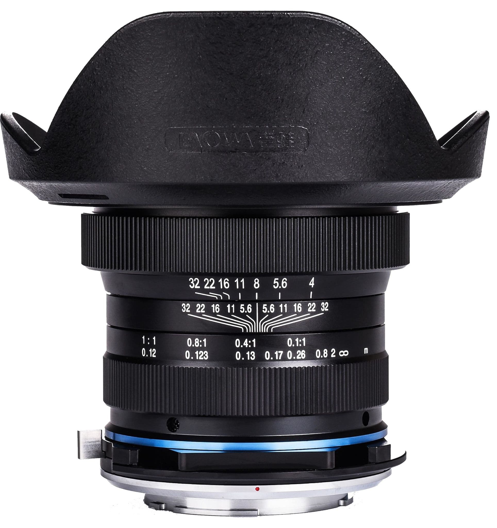
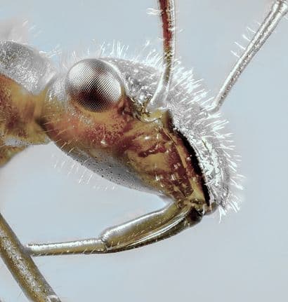
Macro Photography
Is the use of specialised lenses for the close up photography of small subjects such as plants, insects and microfossils. Typically a macro lens will reproduce images of subjects at a ratio of 1:1 (and sometimes greater) with respect to the camera’s sensor size. This is sometimes called ‘life-size’ photography. When printed or observed on a screen the subject is therefore significantly magnified.

Ultra Macro Photography
Is the use of specialised lenses for the ultra close up photography of small subjects such as parts of features of plan leaves and flowers, small insects and insect parts, fungi and so much more. Typically an ultra macro lens will reproduce images of subjects at a magnification far greater than 1:1, (eg 1X to 5X), with respect to the camera’s sensor size. When printed or observed on a screen the subject is therefore very highly magnified. Working at these magnifications overlaps the magnifications achievable with micro photography (or photomicrography if you prefer).
Example macro and ultramacro lenses can be seen HERE
TIPS
Stability is crucial use of a cable shutter release is highly recommended to avoid blurred images created by camera shake, the latest generation of cameras that do not have mechanical shutters are beneficial in this respect.
Also a decent quality, sturdy tripod is recommended when in the field or studio.
Inevitably at high magnifications the depth of focus can be rather narrow, it is also affected by the shutter setting selected. Many macro photographers therefore use focus stacking software and focus control hardware with impressive effect to give effectively an infinite depth of focus without compromising sharpness.
Illumination can also be an issue when close to the subject and illumination diffusers can help a great deal with high reflective subjects.
FIELDWORK
Macro photography can be used in the field very successfully if the subject is not moving, photographing moving insects and plants on outside is very much more of a challenge. Fine XY controls on the camera mount can be very helpful for subject framing, A manual XY focussing rail is ideal. There are some handy flexible specimen holders which can stabilise plants. Illumination is highly critical and some good portable lights can make the difference between an average and an exquisite shot. It is also possible to use a motorised macro rail with a battery pack for rapid sequence capture for focus stacking.
Studio work is simpler to do as the lighting, camera stability, camera positioning, angles etc. can be much more easily controlled. The use of angled viewfinder screens, angled viewfinder eyepieces and field monitor screens can all make setting up for that perfect macro shot so much easier.
MACRO & ULTRA MACRO LENSES
Macro lenses are typically long barrel lenses for close-up work. They can have a variety of focal lengths depending on the type of work being tackled for instance:
90-105mm macro lenses
Favourites of many because they are ideal for photographing small subjects such as plants and insects at a reasonably comfortable distance and the depth of focus is relatively deep.
150mm – 200mm macro lenses
For additional working distance then you need 150mm – 200mm macro lenses
45-65mm macro lenses
For very close-up work providing you can control the positioning and environment more rigorously and where you require a natural background perspective 45-65mm lenses can be used
Variable focal length macro lenses
Of course there are also continuously variable focal length lenses which can be adapted to almost any macro photography situation.
CONVERTING LENSES TO BECOME MACRO LENSES
It is possible to convert lenses to become macro lenses by adding extension tube or bellows alternatively by adding a close-up lens (also called filter) to the front of the camera’s lens.
Extension Tubes
Extension Tube examples are 12mm, 25mm, 34mm etc often supplied in sets of 3 an there are adjustable versions. These are relatively low cost hollow tubes that simply space the lens from the sensor. A low cost alternative to dedicated macro lenses and very good for experimenting but the lens aperture information may be lost. These are not necessarily the best optical solution as they will magnify any optical aberrations in the lens if it is not optimised for close distance work and field flatness. There is also potential for light leaks, you may need to use black tape to seal it up. Internal reflections can be an issue in poorly-baffled tubes requiring matt black flock tape inside the tube.
Bellows
Not so common these days the effect is the same as extension tubes Having an advantage over extension tubes in that the extension is variable. Not commonly available and they are more complicated to mount and to use.
Reversing the lens with a reversing kit
Another technique is to reverse a lens using a reversing ring which you attach to the filter thread on the front of the lens allowing you to attach it in reverse. A very cheap and often effective technique being deal for lenses below 50mm and make sure the lens has a manual aperture setting so you can adjust it. Just attach to the filter thread of the lens, turn it all around and re-attach in reverse. This can be used with extension tubes for extra magnification. You may lose the ability to focus at infinity and the lens aperture information will be lost.
Close-Up lenses
Also known as filters and enlarger lenses - the most popular are 40mm or 50mm, they were originally designed for photographic enlarging of a negative onto printing paper. When used they give exceptional flat field imaging at a very reasonable cost. You can use these in reverse but it must be an asymmetric lens for best results and it should be better when used reversed but generally below 50mm. Be aware of potential light leakage due to a window fo. digit illumination – reversing the lens eliminates this Some adapters can be difficult to find requiring typically a M39 adapter – a simple step ring is easier to find if used in reverse, it is best to buy a 6 element achromatic lens
OTHER USEFUL KIT for Macro and Ultra Macro Photography
IRIS

M42 Iris for depth of focus extension with extension tubes and bellows
ILLUMINATION

Flash Bracket
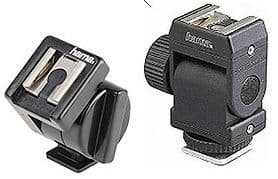
Adjustable Flash Shoe Mount for horizontal mounting

Ringlights and Diffuse Lights
SPECIMEN HOLDERS & CLAMPS


Clamps and Clips


XY Stages & Stands for holding, illuminating and moving subjects under fine control.
TRIPODS

Solid, heavy ideally – stability is king
MACROBEAN BAG

Great when a tripod simply isn't convenient.
XY CONTROLS

for fine specimen control
FOCUS RACKS & RAILS

Tripod mounted

Bench Mounted

Motorised
ANGLED EYEPIECE VIEWFINDER

Allows you to view from above when your camera is at different angles. Makes focussing much easier if you do not have the latest adjustable viewfinder
FIELD KITS

Battery packs for motorised focus rails

Flexible specimen clamps - stops things moving in the wind.
FOCUS STACKING

Software to create fully focussed images from a stack of partially focussed high magnification images
Micro Photography
Microphotography can be achieved in one of two ways using a DSLR camera:
- Adding a microscope lens (objective) to your camera or lens system we call this Objective Micro Photography, click on the link to learn more
- Mounting your camera onto a microscope
- See here for some useful definitions and explanations
DEFINITIONS & EXPLANATIONS
Micro Photography (otherwise known as photomicrography)
For the simple expedient that micro photography is much easier to say that photomicrography and conveys, for most, exactly the same meaning, we will refer to it as micro photography.
Micro photography is the taking of pictures (and videos) of subjects at higher magnification than can be produced by macro and ultra macro lenses. In practice that means magnifications ranging from 2x to 1200X (the theoretical maximum for optical microscopy). However, the vast majority of amateur microscopy rarely exceeds 100X.
WHAT DOES MAGNIFICATION MEAN?
Much more complicated than you think!
This is the change in size from the actual subject size to the size projected. Because the word 'projected' means different things to different users the stated value of magnifications can be extremely misleading.
OPTICAL MAGNIFICATION
For instance, looking through an eyepiece on a microscope the image is projected onto your pupil, these are pretty constant in size so magnifications given by a fixed optical system of say 100X to your pupil (made up from say a 10x eyepiece and a 10x objective multiplied together), are simple to understand. This can be referred to as the optical magnification.
CAMERA IMAGE CAPTURE MAGNIFICATION
However, when a DSLR is attached, magnification now becomes the size of the subject relative to the size of the camera sensor. The sensor is usually bigger than your pupil. So with exactly the same optical system as above the magnification becomes 80X perhaps.
DISPLAY MAGNIFICATION
But what if you are observing the image on a screen, effectively using a digital microscope, well then , it is utterly confusingly argued the magnification is relative to the size of your screen, so the same optical system as above could easily be quoted as 1000X! Digital microscope companies would even say that the magnification doubles if you simply observe the image with a 2X bigger screen. The magnifications given by digital microscope manufacturers are nearly always wholly misleading and, as larger magnifications attract more customers and as many digital microscope manufacturers are essentially low price toy manufacturers with no grasp of (nor regard to) the fundamentals, these high magnification values are relentlessly advertised - beware.
Clearly this is all completely confusing but all correct at the same time!
There is only one value that matters and that is the diameter of the observed field of view. ( Eg 1mm) Unfortunately this is the only number that is extremely rarely given. It is wisest therefore to try to find the optical magnification (ie the magnification to a pupil), which is always stated with respect to optics but almost never with respect to digital microscopes.
WHAT MAGNIFICATION SHOULD I USE?
As in macro photography, the higher the magnification you use, the narrower is the depth of focus, the more difficult is it to mount and illuminate the subject and the more important it is to have everything stable and under fine control.
Here is what scientists generally use (examples):
Most professional scientists and staff in inspection laboratories in industry looking at lumpy and bigger subjects such as undissected plants, electronic components, fabrics, print quality, particulates, concrete, fossils, insects, waterborne insects etc usual are looking at subjects that are not much bigger than 10-20mm. They may well want to zoom in a bit and see field of view about 2 mm across. So optical magnifications of 5X-50X are all that is needed. These magnifications are provided usually by a stereo zoom microscope, a monozoom microscope can also do this - more about these choices later.
Overwhelmingly the stereo microscope is the most popular (and suitable) type of microscope for the amateur microscopist.
Scientists studying prepared material such as thin sections of tissue (eg histology), polished sections through metals (eg metallurgy), stained fungal spores, and parts of dissected organs (eg physiology, entomology and zoology) do so with higher optical magnification microscopes such as upright biological or metallurgical microscopes. Optical magnifications of 40X-1000X are used.
Scientists looking at unstained subjects such as pollen, waterborne invertebrates, certain blood cells (eg haematology) and cells in culture (eg cytology) use biological upright microscopes at higher optical magnifications but with special techniques such as phase contrast and differential interference contrast (DIC). Optical magnifications of 40X-1000X are used.
Scientists looking at prepared, stained, thin sections of tissue of fluorescent stained cells use biological upright microscopes at higher optical magnifications sometimes with fluorescence outfits. Optical magnifications of 40X-1000X are used.
See our Choosing a Microscope Technical Tips
WHAT METHODS CAN I USE FOR MICRO PHOTOGRAPHY?
OBJECTIVE MICRO PHOTOGRAPHY
Attaching microscope objectives (lenses) direct to your DSLR camera using special objective tube kits. Essentially you are building your own microscope. To do this you take an objective that is normally found on a biological or materials upright or inverted microscope and attach it to your DSLR camera in one of two ways:
Attach to the end of your telephoto lens. To do this you need a 42mm:objective thread adapter. Most objectives have what is known as an RMS thread, others have M26 threads and very occasionally other size threads. So you will need the correct M42 adapter. For this method, ideally you will need an infinity corrected objective (see below).
Attach to an M42 Extension Tube Set. This set starts with a bayonet mount:M42 adapter that is specific to your camera. Then there are a number of extension tubes so you have the correct length. Finally you will need the 42mm:objective thread adapter outline above. The exact length of the tube set depends on the 'tube length' stated on your microscope objective, this will require some experimentation to optimise. Please see below:


About Objectives
MAGNIFICATION
Lower magnification objectives below 4X , counter-intuitively, usually cost much more than high magnification ones, this is because they are rarer and require a lot of engineering to work well on a microscope. Lower magnification objectives, as in macro photography, are always easier to use as they generally have a longer working distance.
QUALITY OF OBJECTIVES
The quality of the optics hugely influences prices as you would expect. In order of increasing quality these are the choices in quality, ranked from best at top:
PLAN APOchromatic
Semi PLAN APOchromatic - sometimes also called PLAN FLUORITE
PLAN achromatic
Semi or Economy PLAN achromatic
Achromatic
WORKING DISTANCE
This is the distance from the end of the objective lens to the point on the subject that is in focus.
You should be aware that the higher the quality of the objective, the shorter is the working distance. Therefore to make long working distance versions of very high quality objectives is a very difficult task which increases the price hugely.
Certain classes of microscope onto which objectives are designed to be mounted require a long working distance, these tend to be inverted microscopes and some materials/metallurgical microscopes. It is not a bad idea to seek out these objectives but they will cost more.
You can increase the 'native working distance of an objective by introducing an M42 Iris, in exactly the same way as an iris is used in macro photography. Closing the iris to increase working distance requires more light of course and the image quality will degrade.
SPECIAL OBJECTIVES
There are a host of different types. Here are some common examples:
Long Working Distance - inverted microscopes and some materials/metallurgical microscopes use these.
Coverslip Corrected - these objectives typically have 0.17 written on them. They are specially corrected for looking through a coverslip (coverglass) with a thickness of 0.17mm which is placed over a specimen on a glass slide. Sometimes they have a different coverslip correction value. Best to avoid objectives with this written on them if possible unless, of course, you are using a coverslip. It is not a disaster to use objectives up to a magnification of 20X without a coverslip on your specimen, beyond that magnification the image degrades increasingly.
FL - these are usually fluorescence grade objectives and may even be Plan Fluorites, they are designed to allow the transmission of a wider range of wavelengths. They are generally very good for photography.
PH/PHP Phase Contrast, these are designed with an annulus built into them which is supposed be aligned with corresponding disks in a biological upright's phase contrast objective to give a particular contrast enhancing effect on 'gelatinous' subjects that otherwise may be substantially transparent. These objectives also work as normal 'brightfield' objectives and are fine for photography.
DIC Differential Interference Contrast, these are similar to phase objectives but use a polarising special effect. They are usually high quality objectives and are very good for photography.
Motorising Microscopes
An optical microscope can be motorised in many ways. The most common requirement among the amateur community is to motorise the focus so that a sequence of partially focussed images taken at different heights can be collected in order for focus stacking to be used.
However, as soon as you go down the path of motorisation, it becomes apparent that quite a number of functions, by absolute necessity, are required to be orchestrated. In this section we will deal with the capture of images using a DSLR type camera rather than a c-mount microscopy camera.
A summary of a typical process is described below:
1) Attach the motor to your microscope
2) Attach your camera to the microscope and focus on a specimen
3) Connect the motor and a controlling computer to the Motor Control Unit. Connect the shutter cable of the camera to the computer (or controller dependant on the system).
4) Determine and state how many focus steps, the direction and magnitude you need the focus motor to make and enter it into the software on the computer
5) Press start and do make sure nothing disturbs the microscope while it is acquiring images
6) Review the images and put them into Focus Stacking software to process
It is a very similar procedure to using a Motorised Macro Rail.
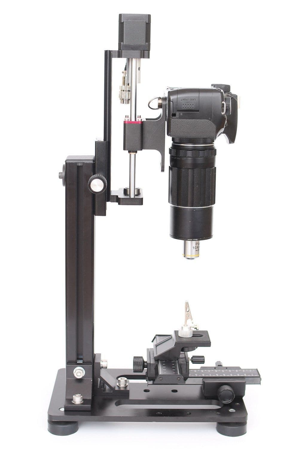
The system we are providing is shown HERE.

The motorised focus system work by attaching to a focus knob on the microscope system that is easily turnable which raises the height of the stage on which the specimen is mount and a focus which does not change position during focussing (some track stands have a system whereby the focus knob moves up or down as the knob is turned). In practise this means that the motor is usually attached to a fine focus knob of a microscope, please note these are rarely found on stereo microscopes (but we can supply) but usually found on biological upright compound microscopes.
Mounting and Presenting your Subjects
For high magnification photography, especially if you intend to do focus stacking, it is important that your subjects are not moving (unless of course you are taking a video sequence).
Ultramacro supply a range of specimen holders, clamps, XY stages and stands to help with this.
See our products HERE
These simple crocodile clips are great for holding subjects either directly or for holding the pins and mounts onto which specimens have been placed such as insects:


You can get fine control of positions with simple versions like these:


These holders can easily be mounted onto stands like this:

There are a range of other useful holders and clamps available:
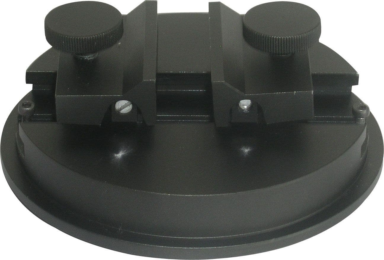

Stages and macro rails are also useful for positioning subjects with fine control over movement:


Focal Lengths and Working Distance in Microscope Optics
Many photographers ask us what is the focal length of a particular microscope objective or a microscope system and how does that differ from working distance (distance from end of lens to the point on the subject that is in focus).
In actual fact calculating the focal length in a microscope system is not particularly useful, it is plainly obvious that the amount of light will be small and simply put the camera attached to Liveview and open any iris and adjust the illumination so you can see what you want, generally we leave the shutter speed on auto.
But if you really want to know, here it is:
Consider the following microscope objective (infinity corrected):
Magnification =50×
Glass Thickness =3.5
mm
NA =0.50
WD =13.89
mm
Focal Length =4
mm
In a single lens the working distance would be the focal length. For compound lenses, like microscope objectives, you have to take into account the full optical system to figure out the working distance. Focal length and working distance are not related in a way that you would expect because the objective is a compound lens. The magnification for microscope objectives is also confusing because to calculate the magnification you must know something about the tube lens in the rest of the system that is used along with the objective.
For example,
Suppose you have two focusing lenses (f1, and f2) separated by a distance d were the first lens is a distance W from the object you are trying to image. The complicating factor is that microscope objectives are designed to be mounted onto a rotating turret of different magnification objectives, when rotated the length of the objective plus the working distance of each objective are carefully calculated to be the same so you do not need to refocus after selecting a different objective. So they fit into a specific space so they have a required parfocal distance PD = d + W (I am assuming thin lenses).
The ray tracing optics for this lens system would be:

This resolves to:

In order for this objective to image properly we set the requirement that M12 = 0. When we are happy that our objective correctly images we can figure out the effective focal length by calculating the M21 element.
Suppose that PD = 45 mm (quite a common value), f1 = 11 mm, f2 = 16.5 mm. These are just theoretical values for this exercise, but PD for microscope objectives is 45 - 60 mm.
From the requirement that M12 = 0 we can calculate W:

so the working distance is 19.15 mm.
Next we can calculate the focal distance of this lens, which we can get from the M21 element:

but focal lengths are usually written like M21=−1/fo
so the focal length of the compound lens system is fo = 110 mm. So, very clearly, compound lenses do not behave like single lenses.
What about the magnification? Here the M matrix is created with all the numbers plugged in:

By looking at this you would expect the magnification of this lens to be 1.35 (the minus sign just means the image is upside down). However, the magnification for microscope objectives require knowledge of the tube lens.
For example stated magnifications are based on a tube lens focal length of 200mm (common for infinity objectives)
So for our example above using a 200 mm tube lens the magnification would be M=ftube/fobjective=200/110=1.82

